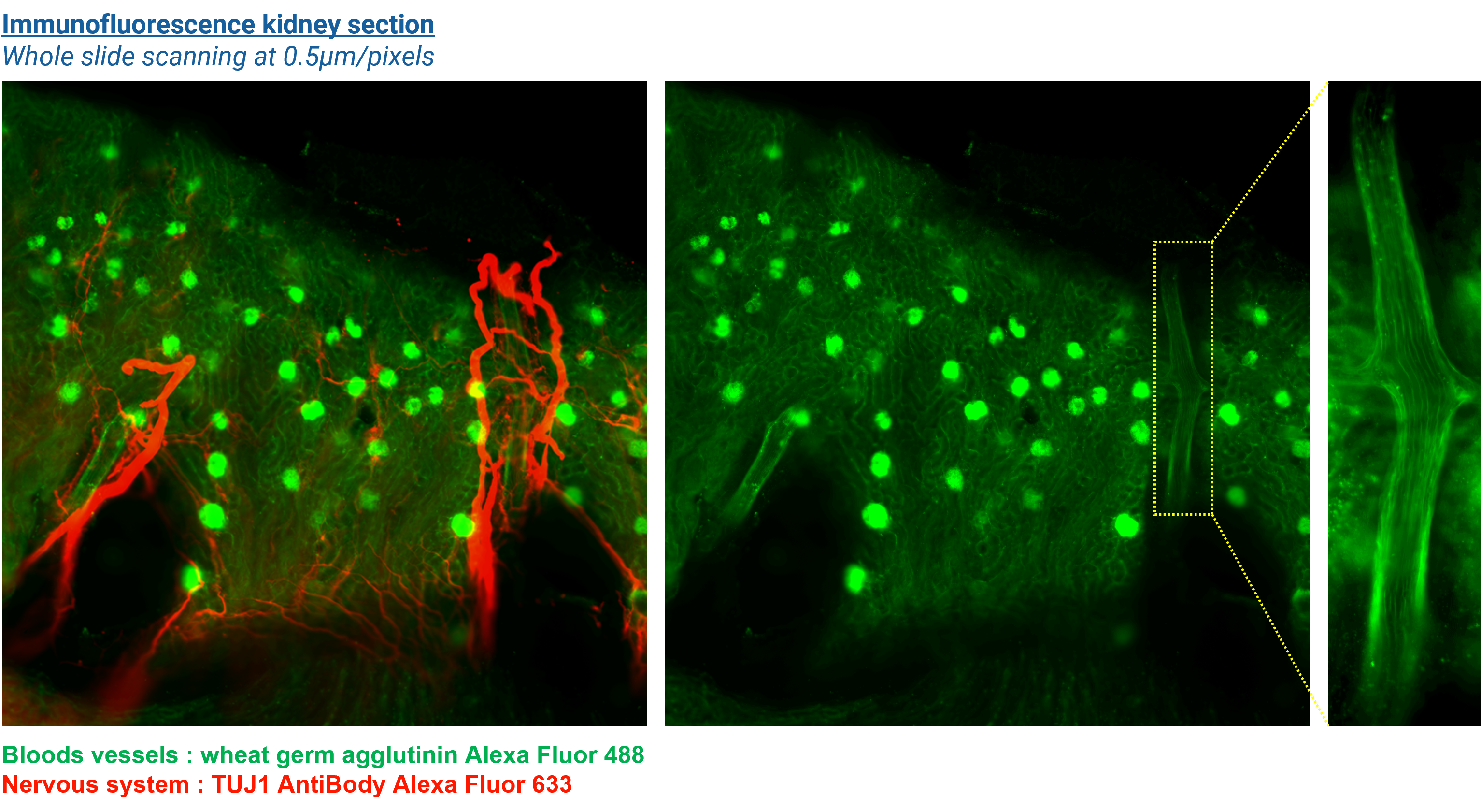
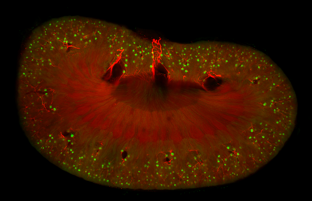
Using fluorescence-labelled antibodies against specific antigens make it possible to highlight the presence of a molecule of interest directly within a tissue section. Therefore, tissue immunofluorescence allows defining the expression of protein biomarkers and their localization within the tissue, leading to topographical mapping of biomarkers.
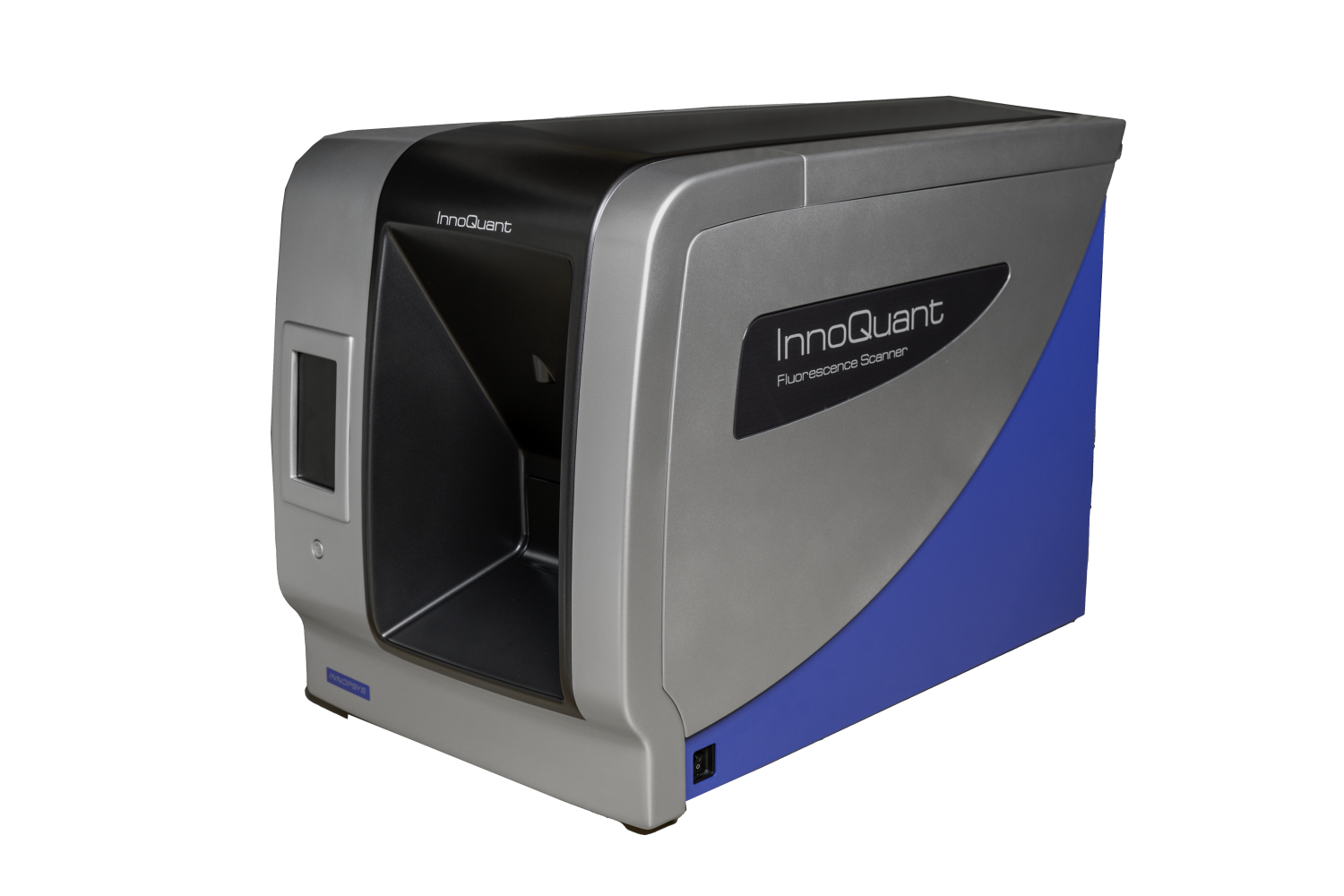
To carry efficient and reliable topographic mapping of biomarkers in tissues, it is necessary to obtain a whole overview of the tissue section. Some motorized microscopes can do this by stitching each individual view captures to rebuild the tissue image. However, the tiling techniques are hard to perform and some errors in stitching can occur. The InnoQuant innovative technology can scan the whole slide without any tiling or stitching processes.
InnoQuant enhances the multiplexing capabilities of fluorescence-based immunostaining. Indeed, the InnoQuant can scan up to 4 fluorescence channels simultaneously. Therefore, the localization of 4 independent biomarkers can be done on a single and intuitive process. The InnoQuant is a powerful tool for large-scale tissue section studies applied to cancer, virology, immunology or neuroscience fields.

Products


